
2 neobiotech IS II Active fixtures were placed in immediate implantation 4 months ago in #24,#25 region.

During 2nd stage surgery, implants were uncovered and attempted to counter torqued to 30ncm.
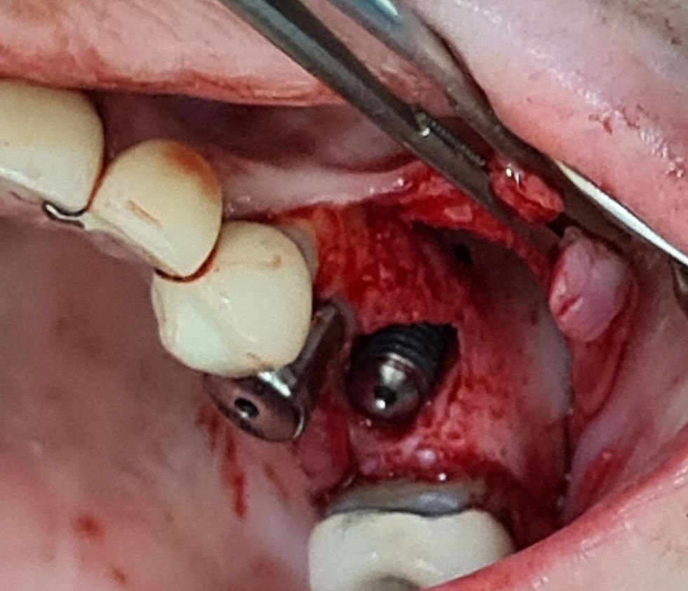
#25 implant was not stable and was easily removed during removal of cover screw.

Note bone attached to fixture at top 1/3 of fixture.
Concluded graft failure due to residual granulation tissue around implant
Site debrided and large socket of healthy bone was created.
Labial fenestration after curettage.

Depth probe from Master KIT is helpful in dragging out granulation tissue from sockets, especially helpful in small drill osteotomy diameters.
As mesial distal bone volume is insufficient, we could not use a larger diameter fixture to engage the socket walls.
Sinus Floor is targeted for apical engagement of fixture to inferior wall of sinus using sinus all kit

Initial preparation with Lindemann drill with drill stoppers deviated.

Osteotomy position corrected and preparation into sinus floor with S reamers


Apical fenestration and socket of implant grafted with allograft